

Late breast cancer relapse may involve slow-growing cancer cells that survive hormone-blocking therapy. Fernandez et al. identified Rac1 signalling as a key escape pathway in these therapy-tolerant cells. Rac1-blocking drugs improved outcomes in preclinical models, while soy isoflavones with Rac1-related activity were linked to 26% lower recurrence risk in a meta-analysis. This highlights Rac1 as a promising relapse-prevention target.
For many women with breast cancer, finishing the first phase of therapy does not always mean the threat is gone for good. This is especially true for oestrogen receptor-positive breast cancer (fuelled by oestrogen), where hormone-blocking therapy can suppress tumour growth. Yet about 20-40% of cancers still return years or even decades later. Why? A major new study suggests that a population of slow-growing cells may survive therapy and gain the ability to spread elsewhere (metastasis) through the P-Rex1/Rac1 signalling pathway. Now, could we target this pathway to reduce the risk of relapse for breast cancer survivors?
Understanding Breast Cancer Relapse
The Slow-Growing Cells That Refuse to Disappear
Oestrogen receptor (ER)-positive breast cancer is often treatable because it has a clear weakness: it depends, at least partly, on oestrogen to grow. This is why many patients take hormone-blocking therapies for five to ten years after initial therapy (usually surgery with radiotherapy and/or chemotherapy) to suppress any remaining cancer cells.
However, this protection is imperfect. About 20-40% of survivors still develop therapy-resistant, metastatic relapse. This means that the original breast cancer has spread to other organs (metastatic) and has become much harder to control (therapy-resistant). At this stage, the disease is considered incurable, with a median survival of around 3 years only.
More concerningly, these relapses can appear very late, sometimes 10 or even 20 years later, when the cancer may seem like a closed chapter. Late relapses are also harder to anticipate because they do not always carry the usual warning signs of aggressive disease. Many existing tests are better at identifying cancers that are likely to return early, especially those with fast-growth signals such as high tumour grade or proliferation markers.
For a long time, one of the main explanations for late relapse of breast cancer has been dormancy. Some cancer cells enter a sleep-like state, remain hidden in the body and start growing again years later. However, dormancy may not always mean complete inactivity. Some cancer cells may continue to grow, albeit at a rate so slow that they remain clinically undetectable. One estimate suggests that the doubling time of breast cancer cells may range from 50 to 400 days. In slow-growing cancers, where the doubling time exceeds 250 days, it may take more than 15 years for the tumour to become large enough to detect.
In a new study published in Nature Communications, Fernandez et al. at the Garvan Institute of Medical Research in Australia explored whether slow-growing cancer cells could drive late metastatic relapse. In ER-positive breast cancer, they first noticed that metastatic tumours had lower levels of Ki67 (a cell proliferation marker) than the original breast tumour. In other words, some metastatic lesions were surprisingly slow-growing. This pattern was not seen in ER-negative breast cancer, suggesting that slow growth may be a distinctive feature of late metastatic relapse in hormone-sensitive breast cancer.
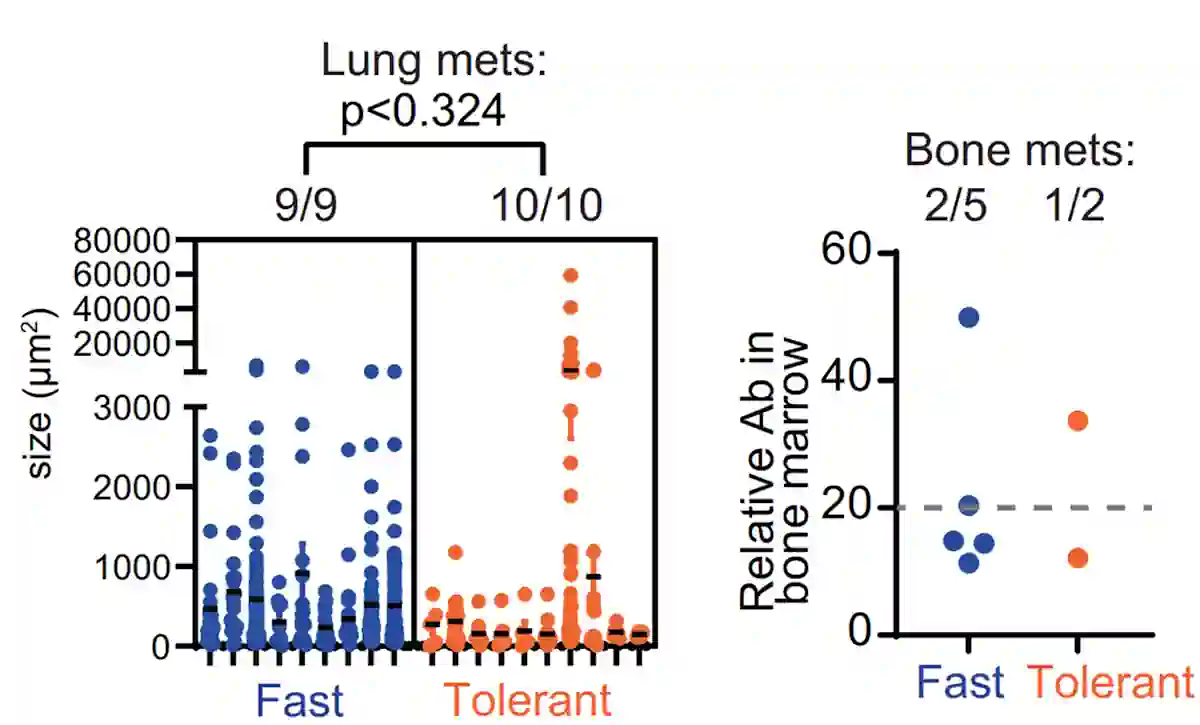
Figure 1. Following hormone-blocking therapy, slow-growing breast cancer cells still retain their metastatic capacity. Fast-growing therapy-resistant breast cancer cells are shown in blue, while slow-growing therapy-tolerant cells are shown in orange. Despite growing more slowly, the therapy-tolerant cells still spread to the lungs, with lung metastases similar in size to those from fast-growing cells. Bone metastases were also detected in a small subset of both groups. Source: Fernandez et al. (2026), Nature Communications.
Under the microscope, these slow-growing tumours also looked unusual. They had denser collagen around them, as well as enlarged or abnormal nuclei. These are actually features of a senescent-like state, i.e., a stressed, ageing-like condition in which cells slow down or partially stop dividing. When the study profiled the gene activity, they found that the slow-growing cancer cells resembled a small subset of cells that appeared shortly after hormone-blocking therapy. This suggests that hormonal therapy may, in some cases, reprogram cancer cells into a slow-growing, therapy-resistant state that still retains metastatic ability.
“It took years to isolate these specific cells because they were dividing so slowly, almost in defiance of how we typically expect cancer to behave. But once we observed them in action, we realised that a slow clock doesn’t mean a stopped clock,” the study lead author, Kristine Fernandez, PhD, said in a press release. “These cells were migrating to organs like the bone and lungs, proving that speed isn’t everything when it comes to metastasis.”
A Possible Weakness: P-Rex1/Rac1 Signalling
Fernandez and colleagues then asked what distinguished these slow-growing cells. They found that fast-growing cells showed higher activity in well-known growth pathways, particularly the epidermal growth factor receptor (EGFR) pathway. In contrast, the slow-growing cells displayed a different pattern, centred on the P-Rex1/Rac1 pathway. In this pathway, P-Rex1 activates Rac1, a protein that helps cells survive, move, and invade other tissues.
When the study downregulated P-Rex1 or blocked Rac1, the slow-growing cells became less migratory. The study also found that prolonged tamoxifen (an oestrogen-blocking drug) exposure increased both P-Rex1 and Rac1 activity in a mouse model of ER-positive breast cancer. Patient data further showed that higher P-Rex1 activity was associated with late relapse, while the broader Rac1 pathway was linked to poorer relapse-free survival.
After identifying P-Rex1/Rac1 signalling as a possible weakness in these slow-growing cells, Fernandez et al. asked whether blocking this pathway could be a viable therapeutic strategy. In laboratory experiments, Rac1-blocking compounds reduced the survival and motility of slow-growing cells more than those of fast-growing cells. The study then tested this idea in mice using a tumour grown from a patient whose ER-positive breast cancer had already spread and had stopped responding to standard therapies. On their own, the Rac1-blocking drugs had only modest effects. However, when combined with tamoxifen, they shrank tumours more effectively than tamoxifen alone (Figure 2). This suggests that blocking Rac1 may help target the slow-growing cells that survive hormone therapy and drive relapse.
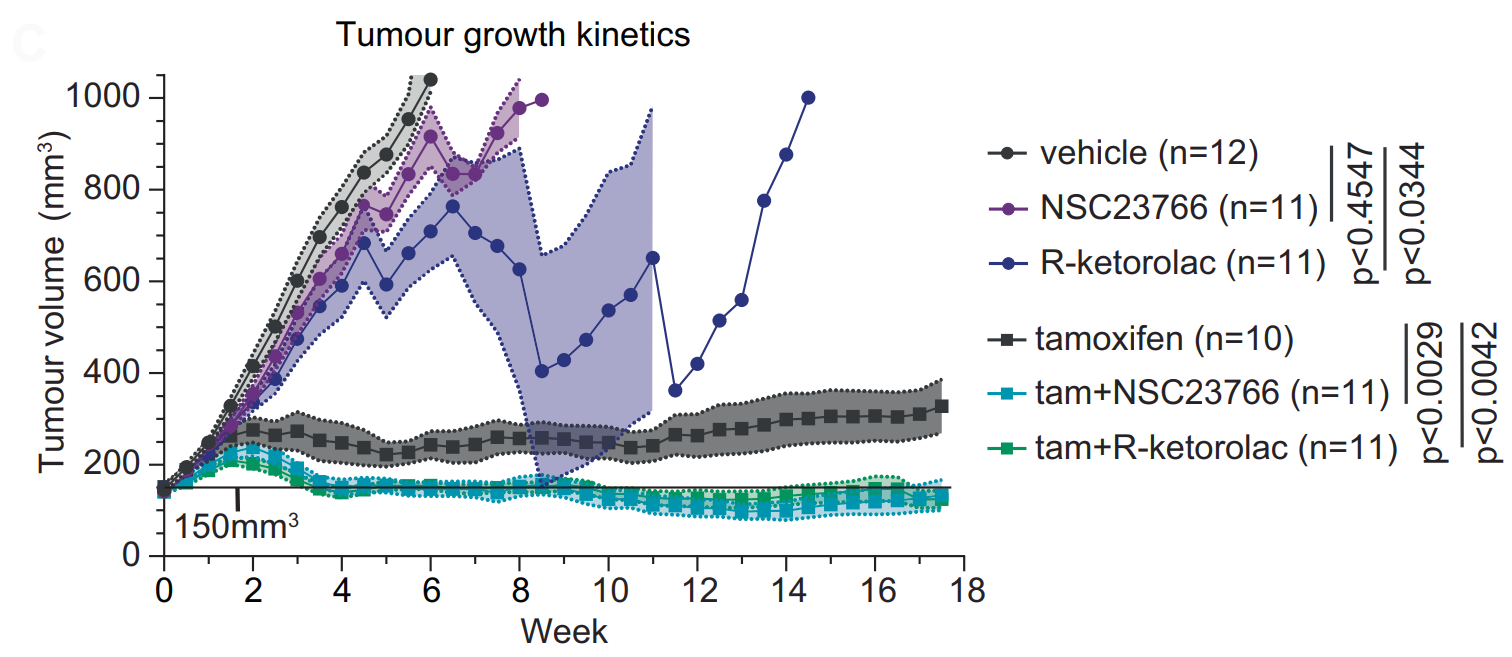
Figure 2. Combining tamoxifen with Rac1-blocking drugs produced the greatest tumour shrinkage. In mice carrying breast tumours derived from a patient whose oestrogen receptor-positive breast cancer had become resistant to standard therapies, tamoxifen alone slowed tumour growth but did not stop it. Rac1-blocking drugs (NSC23766 or R-ketorolac) alone had modest effects. However, pairing tamoxifen with a Rac1 blocker resulted in stronger, more sustained tumour shrinkage. Source: Fernandez et al. (2026), Nature Communications.
Finally, the study looked at previous clinical data on ketorolac, a painkiller sometimes used during breast cancer surgery. Why ketorolac, though? Unlike other painkillers, ketorolac has an off-target Rac1-inhibiting activity. Across several cohorts, patients who received ketorolac had a lower risk of breast cancer relapse – a pattern not seen with other painkillers without a known Rac1-related activity (Figure 3). While this intriguing finding does not yet prove that ketorolac prevents relapse, it warrants future clinical trials testing whether ketorolac or Rac1-targeting compounds could reduce relapse risk in breast cancer.
“For a long time, the idea that extremely slow-growing cells could drive relapse was just a theory. We’ve found evidence for the way this could happen in ER-positive breast cancer,” the study’s director, Associate Professor Elizabeth Caldon, PhD, said in a press release. “By identifying the pathways that are important in these slow-growing cells, we have a new lever to potentially prevent these deadly outcomes.”
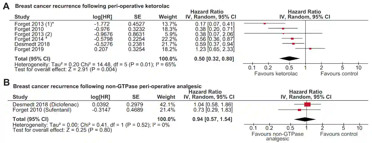
Figure 3. Ketorolac, but not other similar painkillers, was associated with a lower risk of breast cancer relapse. This meta-analysis combined results from prior observational studies of women who received pain relief around the time of breast cancer surgery. A hazard ratio (HR) below 1.0 indicates a lower risk of relapse, whereas an HR above 1.0 indicates a higher risk. Overall, patients who received ketorolac had about a 50% lower risk of breast cancer relapse (HR = 0.50), whereas other painkillers without known Rac1-related activity showed no clear association (HR = 0.94). Source: Fernandez et al. (2026), Nature Communications.
Could We Target Rac1 Signalling with Natural Compounds?
Interestingly, several natural compounds have shown preclinical evidence of targeting Rac1 activity and its related processes in cancer cell movement and invasion. Preclinical evidence refers to studies done with laboratory-cultured cells or animals, so these findings are usually considered preliminary. Some examples of these natural compounds are:
- Epigallocatechin gallate (EGCG): As the main catechin in green tea, EGCG has been shown to reduce the migration and invasion of breast cancer cells by lowering the activity of both Rac1 and VASP, another protein involved in cell movement.
- Reishi mushroom: In breast cancer cells, Reishi extract reduced cell movement and invasion by dampening Rac1-related signalling. It also disrupted lamellipodia, the thin, sheet-like extensions that cancer cells use to crawl through tissue.
- Curcumin: In a cell model of metastatic breast cancer, curcumin extract reduced the migration of cancer cells and the expression of Rac-1 and MMP-9. MMP-9 helps cancer cells degrade surrounding tissue, while Rac1 coordinates cell motility.
- Others: Genistein and quercetin may also affect Rac1-related migration pathways, although the evidence is mainly from other cancer models. In brain cancer cells, genistein reduced their migration and invasion by blocking an upstream pathway that activates Rac1, while quercetin inhibited Rac1 activation more directly.
Population studies add another layer of evidence. Some plant compounds that affect Rac1-related signalling in the laboratory are also linked to better breast cancer outcomes in humans, although these studies cannot prove that Rac1 was the mechanism.
The clearest example is soy isoflavones, including genistein. A 2023 meta-analysis of breast cancer survivors found that higher soy isoflavone intake was associated with a 26% lower risk of relapse, especially among survivors of ER-positive breast cancer. The greatest reduction in risk was seen at around 60 mg/day, with no added benefit at higher intakes (Figure 4). Green tea is another example: Meta-analytic evidence reported that higher green tea intake is also associated with a lower risk of relapse in early-stage breast cancer.
All things considered, natural compounds may have a role in supporting the long-term survival of breast cancer patients by limiting the ability of surviving cancer cells to move and spread. The lesson is not that natural compounds can replace standard cancer therapy. It is that natural compounds and Rac1-blocking drugs may deserve closer attention as possible tools to address one of oncology’s most persistent problems: late metastatic relapse of breast cancer.
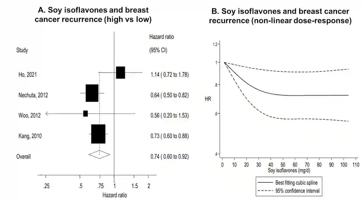
Figure 4. Higher soy isoflavone intake was associated with lower breast cancer recurrence risk. (A) Pooled observational studies comparing women with higher versus lower soy isoflavone intake. Overall, higher intake was linked to a 26% lower risk of recurrence (HR = 0.74). (B) The dose-response pattern showed that relapse risk decreased as soy isoflavone intake increased, then levelled off at around 50–60 mg/day. Source: Diana van Die et al. (2023), Journal of the National Cancer Institute (JNCI) Cancer Spectrum.